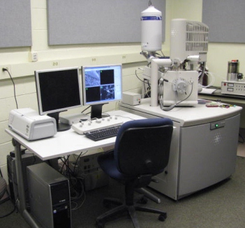
FEI Quanta 200 FEG MKII scanning electron microscope

Traditionally SEM images are acquired by detecting the emission of secondary electrons. However this is only a small part of the information to be gained from the specimen due to its interaction with the primary electron beam. This facility's SEM also has the capability of detecting backscattered electrons (BSE) and electromagnetic radiation in the form of x-rays (x-ray microanalysis).
Our FEI Quanta 200 FEG MKII scanning electron microscope (SEM) was installed in the Spring of 2007. It is a high resolution environmental microscope (ESEM) capable of running in high vacuum, variable pressure and environmental modes which means that it can handle all specimens even uncoated, non-conductive samples as well as wet samples that require being above the vapor pressure of water. It has 1.5 nm resolution, high output thermal field emission (> 100nA beam current) microscope with a high sensitivity (18 mm) backscatter (BSE) detector for atomic number contrast. It also is equipped with an Oxford –Link Inca 350 x-ray spectrometer with a 30 sq mm window for light element diction and spectral imaging and phase analysis.